New multi-functional cardiac imaging capability will benefit range of patients, including older adults and those living with renal conditions, among others
.jpeg?sfvrsn=5f25f2c5_1)
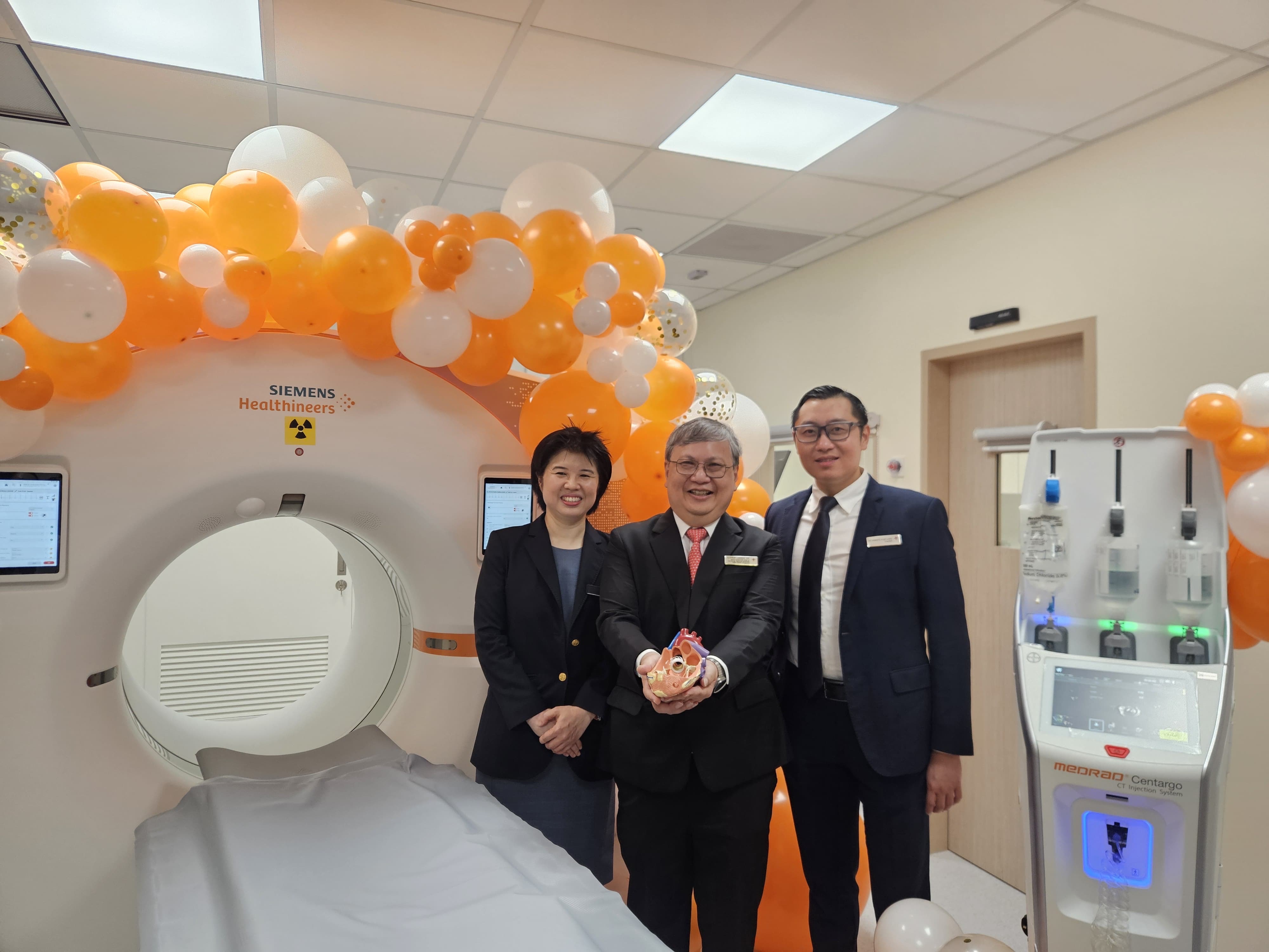
The National University Heart Centre, Singapore (NUHCS) is the first tertiary centre in Singapore to deploy the Photon-Counting Computed Tomography (PCCT) capability for a high workload of complex cardiac cases. Photo credit: National University Health System (NUHS)
(From left to right) Mr Steven Bell, Senior Vice-President, Diagnostic Imaging and Advanced Therapies, Asia Pacific and Japan, Siemens Healthineers; Adjunct Professor Mark Edward Puhaindran, Chairman, Medical Board, National University Hospital (NUH); Professor Aymeric Lim, Chief Executive Officer, NUH; Associate Professor James Yip, Executive Director and Senior Consultant, NUHCS; Professor Khong Pek Lan, Head and Senior Consultant, Department of Diagnostic Imaging, NUH; Adjunct Associate Professor Lynette Teo, Senior Consultant, Department of Diagnostic Imaging, NUH.
SINGAPORE — The National University Heart Centre, Singapore (NUHCS) is the first tertiary centre in Singapore to deploy a new generation of cardiac imaging technology – known as the Photon-Counting Computed Tomography (PCCT) – for a high workload of complex cardiac cases.
As Singapore’s population ages and the burden of heart disease continues to rise, the need for safer, more efficient cardiac diagnostics has never been greater. By 2030, nearly one in four Singaporeans will be aged 65 or older, and cardiovascular disease already accounts for nearly one in three deaths in the country.
In response to this evolving need, the NUHCS and the Department of Diagnostic Imaging at the National University Hospital (NUH) are strengthening care for high-risk cardiovascular patients with the PCCT. This introduces a new approach to imaging for these patients who might previously have to undergo an invasive coronary angiogram1 for cardiologists to visualise their coronary anatomy, before diagnosing and treating their condition.
“With Singapore’s ageing population, we are seeing more patients with complex cardiac conditions who may not tolerate invasive coronary angiogram well,” said Associate Professor James Yip, Executive Director and Senior Consultant, NUHCS.
“Our priority is to continually evolve how we deliver care – making it safer, more precise, and less burdensome for patients. This advancement allows us to reduce reliance on invasive diagnostics while maintaining a high level of clinical confidence, ultimately improving patient outcomes and experience.”
Patient-centred cardiac care
A 90-year-old man with severe aortic stenosis2 was the first patient from NUHCS to benefit from this new capability, undergoing evaluation and subsequently treatment using this advanced imaging approach.
A Transcatheter Aortic Valve Implantation (TAVI) is a common minimally invasive treatment option that replaces the narrowed aortic valve without open-heart surgery. Traditionally, patients undergoing TAVI may require both a computed tomography (CT) scan and an invasive coronary angiogram to assess their heart condition adequately pre-procedure. However, invasive angiography carries additional risks, discomfort, and recovery time, particularly for older patients. It also typically requires higher doses of contrast media, which can pose further risks to the kidney function for those already suffering from renal disease. In Singapore, kidney disease affects more than 500,000 people.
With the introduction of the next generation PCCT scanner, clinicians were able to obtain clear and detailed images of the patient’s coronary arteries using a single scan, avoiding the need for the invasive procedure. This allowed the team to minimise the use of contrast media as well, which was a key clinical consideration as the patient also had underlying kidney issues.
The 90-year-old patient’s care team comprising (from left to right) Adjunct Associate Professor Lynette Teo, Senior Consultant, Department of Diagnostic Imaging, National University Hospital (NUH); Associate Professor James Yip, Executive Director and Senior Consultant, National University Heart Centre, Singapore (NUHCS); and Dr Ivandito Kuntjoro, Senior Consultant, Department of Cardiology, NUHCS. Photo credit: National University Health System (NUHS)
Professor Khong Pek Lan, Head and Senior Consultant, Department of Diagnostic Imaging, NUH, shared: “Many cardiac patients come in with conditions that result in fast heart rates, heavy calcification, or with existing stents that can make it challenging to obtain clear, reliable images. With this new capability, we can visualise coronary arteries and cardiac structures in a single CT scan with much greater clarity, even in difficult cases, while using less contrast media. This enables us to make confident decisions while offering high-risk patients such as older adults and those with kidney disease a safer diagnostic journey.”
The patient subsequently underwent a smooth TAVI procedure and was discharged without complications after two days. Post-procedure assessments showed stable and improved kidney function, and no additional interventions were required.
Advances that meet evolving healthcare needs
This approach ultimately reflects a broader move towards less invasive, more patient-centred cardiac care with tangible benefits, including:
- Avoiding unnecessary invasive procedures, which reduces procedural risks, discomfort, and hospital stays
- Lower contrast media usage, which improves safety for patients with renal impairment
- Clearer imaging in complex cardiac cases, including patients with calcified arteries or prior stents
- Consistent diagnostic quality in patients where optimal heart-rate control is difficult
As Singapore’s healthcare system adapts to the demands of an ageing society, these improvements translate into a safer, smoother, and more efficient care experience. The benefits are also applicable to wider populations, including paediatric patients. Ultimately, these developments underscore NUHCS and NUH’s commitment to delivering the best in cardiovascular care, and to continually advance its capabilities to meet Singapore’s evolving healthcare needs.
To download the PDF version of the media release, click here.
1A coronary angiogram is an invasive diagnostic imaging test that uses contrast media and X‑rays to visualise the coronary arteries and detect any narrowing or blockages in blood flow to the heart.
2Aortic stenosis occurs when the aortic valve becomes narrowed and stiff, making it harder for blood to flow from the heart to the rest of the body. This forces the heart to work harder and in severe cases can lead to symptoms such as breathlessness, chest pain, fainting, and heart failure.