If there are signs and symptoms of Sarcoma, your doctor may order some imaging tests. These include, plain X-rays, CT scans, ultrasound scans, MRI scans or even PET or bone scans. The test selected depends on the location of the lump.
After the imaging tests are done, a biopsy is needed to confirm the diagnosis of Sarcoma. This can either be done as a needle biopsy, or a surgical biopsy.
Needle biopsies are done using a core biopsy needle. These remove a 1mm core of tissue from the tumour, and can be done in the clinic or under by a radiologist in the radiology centre using imaging guidance. Multiple cores are often needed in order to obtain sufficient tissue for a diagnosis. Fine Needle Aspiration (FNA) biopsies are not used frequently for sarcomas because insufficient tissue is obtained.
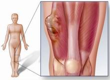
Surgical biopsiesmay be required for some tumours, especially bone sarcomas and large soft tissue Sarcomas.
Biopsies have to properly planned and should be performed by teams with experience in Sarcoma treatment. An improperly performed biopsy may make subsequent removal difficult and may compromise outcomes of treatment.
The specimen taken during the biopsy is processed and reviewed by the pathologist. At times, further these using molecular tests may be required in order to establish the diagnosis.
Once a diagnosis of Sarcoma is made, further imaging investigations may be required to complete the staging of the cancer. This helps to assess the prognosis for the patient.